
Oral examination of the horse's mouth has historically been performed with a long-handled dental mirror. While this method remains valid, endoscopic visualization of the oral cavity has significant advantages, including:
- Visualization of the teeth on a tablet or monitor
- The capture of still and video images
- Improved client communication and documentation in medical records
Equipment
Until recently, the rigid endoscopes available for examination of the horse's oral cavity were costly (in the neighborhood of $6,000-10,000) and the images were of poor quality. However, Rob Pascoe, BVSc, BAEDT, Dipl.AVDC Eq., MRCVS came up with an ingenious and inexpensive ($2000-2500) method to construct an oral endoscopy system. The components of this system are:
- Reconditioned rigid arthroscope, 10 mm diameter, 45 degree lens. These are available on eBay or from Endoscopy Support Services.

- C-mount 45 mm video coupler and 4/3 adapter (Endoscopy Support Services)
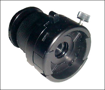
- Panasonic DMC GF6 camera (WiFi enabled, without lens)
- iPad or Android tablet with Panasonic app (free download).
- Portable LED light source, various models are available in the $200-800 range.

In this photo the Cornell veterinary student on the left is operating the endoscope, which is transmitting the image by WiFi to the iPad held by the instructor, Edward Earley, DVM, FAVD/Eq., Dipl.AVDC/Eq. Note that this system is completely wireless and offers real time video display that the owner can watch during the examination. The app allows the assistant to operate the camera from the iPad/tablet; video recording and capturing of still images can be performed by the assistant. Dictation of the examination during the video capture allows later review of the examination and facilitates completion of the patients dental chart.
Here is an example:

This 7 year old warmblood gelding presented with right facial swelling and a right mucopurulent, malodorous nasal discharge. Note that on the endoscopic examination there are defects in the irregular secondary dentin overlying all of the pulp horns of the right maxillary 1st molar (109). Radiographs confirmed apical abscessation of 109.

Treatment consisted of oral extraction and lavage of the right maxillary sinuses for 5 days.
For those of us that have incorporated oral endoscopy into our practice of veterinary dentistry, we can't imagine doing without it. I routinely use it for every oral examination. Please feel free to contact me if you have any questions.
No comments:
Post a Comment