Sunday, December 21, 2014

The Comprehensive Oral Health Assessment and Treatment (COHAT)


In veterinary dentistry, our patients need to be under general anesthesia for a comprehensive oral examination.  This examination involves:

  • An overall assessment of the head for asymmetry, swellings, areas of sensitivity or pain, lymph node or salivary gland enlargement and malocclusion (abnormal bite)
  • An assessment of all soft tissue structures of the oral cavity (tongue, palate, oral mucosa of the cheeks and pharynx, salivary glands, tonsils and laryngeal structures
  • A tooth-by-tooth examination with a periodontal probe and dental explorer; the findings are recorded in detail on a dental chart.
  • Whole mouth intraoral radiographs for assessment of the subgingival dental anatomy and staging of periodontal disease.

Anesthesia

While anesthesia-free veterinary dentistry (equivalent to human dentistry) would be nice, it simply is not possible in small animals, and is not recommended by the American Veterinary Dental College (AVDC). Therefore, it is imperative that veterinary dentists have excellent training in anesthetic techniques and that their operatories are equipped with the best equipment. Many of the patients presented to the veterinary dentist are geriatric individuals, often with concomitant medical problems. Therefore the decision to perform lengthy oral surgical procedures is not made lightly and anesthesia and pain management are as important as the oral surgery itself.

Pre-anesthetic considerations

All patients are given a general physical examination and medical records from the referring veterinarian  are reviewed. Blood is drawn for a complete blood count (CBC) and serum chemistry profile (this is an assessment of liver and kidney function). Dogs with a history of heart disease or a heart murmur may also get a cardiac ultrasound examination. The patients are usually fasted for 8-12 hours prior to general anesthesia.

Multimodal anesthesia

The use of multiple sedatives, analgesics and anesthetic agents that act synergistically, along with the use of regional nerve blocks, allows the clinician to use lower doses of these agents to obtain the desired level of anesthesia. This is very important, in that all anesthetics have potential deleterious side effects that are dose-related. Using smaller amounts of several drugs is much safer than using a higher dose of a single agent to obtain the same level of anesthesia. Equally important is the use of local anesthesia, which also permits the use of much lower levels of gas anesthesia throughout the procedure and allows the patient to recover from anesthesia pain-free.

Patient monitoring and support

Sophisticated monitoring equipment is required for proper monitoring of the patient during anesthesia. The patient's blood pressure, electrocardiogram (ECG), arterial oxygen saturation and carbon dioxide levels and the core body temperature are continuously monitored and recorded by the nurse anesthetist during the surgical procedure.  
Most surgical patients have a tendency to develop hypothermia during the surgical procedure, and this is even more likely in the smaller dogs and cats. To maintain body temperature, which is critical to proper heart function, a heated surgical table and a circulating hot air blanket are used for all dentistry procedures.
Anesthetic agents have some cardiac and respiratory depressant effects. To maintain proper tissue perfusion, the blood pressure is carefully monitored. Any fall in blood pressure is counteracted by lowering the anesthetic gas concentration, increasing the intravenous fluid rate and the administration of medications that improve peripheral blood pressure. Most dentistry patients are placed on a ventilator during the anesthetic period; this insures proper oxygenation of the tissues and prevents elevated concentrations of carbon dioxide in the blood. 

It is easy to see why proper anesthesia adds significantly to the expense of oral surgery in small animals. 

The oral examination

The oral examination usually begins in the conscious patient in the exam room. A slow, quiet
approach will often be well-tolerated by the patient, and an initial assessment of the head and oral cavity can be made. Oral masses, facial swellings, enlarged lymph nodes and fractured teeth can often be appreciated in the conscious patient. This allows the general practitioner to make a tentative diagnosis and leads him/her to a discussion of further treatment options that involve general anesthesia.

Soft tissue structures

With the patient under general anesthesia, a thorough examination of the oral cavity becomes possible. The oral mucosa, the epithelial lining of the entire oral cavity, is examined along with all aspects of the tongue, pharynx and larynx including the epiglottis, vocal folds, tonsils and salivary glands.

Toot-by-tooth examination and charting

Every tooth is examined with magnifying loops and the findings are recorded on a dental chart. Abnormalities of the crown include caries (cavities colonized by bacteria), fractures and developmental defects in the amount or quality of the enamel. The periodontium is the group of four tissues that anchor the tooth to the jaw and includes the gingiva, the periodontal ligament, the cementum and the alveolar bone forming the tooth socket. Periodontal disease is detected clinically by the loss of this attachment that results in deep periodontal pockets, gingival inflammation and recession, and mobility of the tooth. Intraoral radiographs are used to evaluate the amount of bone loss that is associated with periodontal disease. All of these findings are used to stage the periodontal disease and make a decision on the appropriate treatment. 
Early periodontal disease (Stage 1) consists of gingivitis, often associated with the accumulation of mineralized plaque, called calculus. This stage of periodontitis is reversed by professional scaling and polishing of the teeth, followed by good at-home oral hygiene by the owners. Malodorous breath (halitosis) is not normal. It is due to the sulfur-containing metabolites of the bacterial biofilm (plaque) on the teeth and calculus. Good oral hygiene in cats and dogs, just as in people, is required  to prevent bad breath.   Stage 2 periodontal disease is defined as loss of less than 25% of normal periodontal attachment. This stage is treated as for stage 1, recognizing that the attachment loss is not reversible. Stage 3 periodontal disease is loss of 25-50% of the normal attachment. Treatment at this stage involves periodontal surgical techniques that are aimed at regeneration of the periodontal tissues and attachment. This is specialized surgery involving the use of bone grafting techniques. Once there has been greater than 50% attachment loss (stage 4), extraction is  the only treatment option.

Intraooral radiography

X-rays of the teeth are absolutely necessary for the complete oral health assessment. Diagnostic quality dental xrays are only obtainable with imaging plates that are placed in the animal's mouth (intraoral). Since this would stimulate chewing in the awake patient, this procedure also requires general anesthesia. Since pathology below the gumline is frequently only evident in x-rays, it is highly recommended that a whole mouth radiographic examination be part of the COHAT. 


Treatment

Treatments of oral disease may be as simple as prophylactic scaling and polishing of the teeth, to as complex as root canal therapy or the surgical treatment of oral cancer. The veterinary dentist is trained in all dental sub-specialties: orthodontics, periodontics, restorative dentistry, maxillofacial surgery, and endodontics. The ultimate goal in veterinary dentistry is to provide the patient with a comfortable, disease-free mouth. Esthetics are of secondary importance in our pets, and are certainly much less important than in human dentistry. However, when possible, veterinary dentists still try to prolong the function of the teeth. 


This dog was presented for re-evaluation of the right mandibular canine tooth which had been fracture 3 years previously and treated endodontically (root canal therapy). Unfortunately, there were six new fractured teeth (files placed in the exposed pulp chambers) that required surgical extractions. Tooth fracture is commonly due to the use of excessively hard chews, such as NylaBones and deer antlers.


This dog had very little calculus or gingivitis (left) but survey intraoral radiographs identified a severely abscessed 2nd molar (right) which was surgically extracted.


 This dog also has minimal gingivitis. However, intraoral radiographs identified a large area of bone lysis, which turned out to be a cyst between the roots of the left maxillary 4th premolar. The cyst lining was carefully curretted and the gingiva was resutured in position.


 This cat has moderate gingival inflammation, plaque and calculus (left). The intraoral radiographs revealed marked tooth resorption of nearly all teeth (right mandibular cheek teeth shown in the xray on the right). Treatment was surgical extraction of all the affected teeth, which left only the 4 canine teeth in this case.


In conclusion, proper dental care requires general anesthesia for a comprehensive oral health assessment and treatment. Since much of the pathology is "below the gumline" and not evident on the oral exam of the awake patient, full mouth intraoral radiographs are routinely obtained.

Tuesday, December 16, 2014

Oral endoscopy in the horse


Oral examination of the horse's mouth has historically been performed with a long-handled dental mirror. While this method remains valid, endoscopic visualization of the oral cavity has significant advantages, including:

  1. Visualization of the teeth on a tablet or monitor
  2. The capture of still and video images
  3. Improved client communication and documentation in medical records

Equipment

Until recently, the rigid endoscopes available for examination of the horse's oral cavity were costly (in the neighborhood of $6,000-10,000) and the images were of poor quality. However, Rob Pascoe, BVSc, BAEDT, Dipl.AVDC Eq., MRCVS came up with an ingenious and inexpensive ($2000-2500) method to construct an oral endoscopy system. The components of this system are:
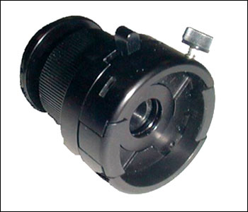
  • Reconditioned rigid arthroscope, 10 mm diameter, 45 degree lens. These are available on eBay or from Endoscopy Support Services

  • C-mount 45 mm video coupler  and 4/3 adapter (Endoscopy Support Services)
  • Panasonic DMC GF6 camera (WiFi enabled, without lens)
  • iPad or Android tablet with Panasonic app (free download).
  • Portable LED light source, various models are available in the $200-800 range.

In this photo the Cornell veterinary student on the left is operating the endoscope, which is transmitting the image by WiFi to the iPad held by the instructor,  Edward Earley, DVM, FAVD/Eq., Dipl.AVDC/Eq.  Note that this system is completely wireless and offers real time video display that the owner can watch during the examination. The app allows the assistant to operate the camera from the iPad/tablet; video recording and capturing of still images can be performed by the assistant. Dictation of the examination during the video capture allows later review of the examination and facilitates completion of the patients dental chart.

Here is an example:


This 7 year old warmblood gelding presented with right facial swelling and a right mucopurulent, malodorous nasal discharge. Note that on the endoscopic examination there are defects in the irregular secondary dentin overlying all of the pulp horns of the right maxillary 1st molar (109). Radiographs confirmed apical abscessation of 109. 


Treatment consisted of oral extraction and lavage of the right maxillary sinuses for 5 days.

For those of us that have incorporated oral endoscopy into our practice of veterinary dentistry, we can't imagine doing without it. I routinely use it for every oral examination. Please feel free to contact me if you have any questions.


Monday, December 15, 2014

Equine Dental Radiography

     Imaging of the equine skull has always been challenging for the equine practitioner. However, with the development of portable digital xray sensors (DR systems), the ability to obtain diagnostic images in the field has greatly improved. This is a brief explanation of the positioning required for diagnostic equine dental imaging and a review of the radiographic anatomy of the skull and radiographic signs of dental disease.


Positioning for equine dental radiographs

I created this paper for the American Veterinary Dental College/Equine Specialty: Dental Radiographic Techniques for the Horse. I also recently presented a short paper at the annual meeting of the American Association of Equine Practitioners (AAEP): How to obtain diagnostic radiographic study of the equine cheek teeth:


During the past year I upgraded my radiography equipment for both small animals and horses with the purchase of Cuattro DR systems for the small animal clinic and the equine ambulatory service. This has made a tremendous difference our ability to consistently obtain diagnostic images. 

Practitioners who would like assistance with obtaining diagnostic images of the skull should feel free to contact me. I would be glad to assist in your interpretation of dental images, which can be sent to my email: rbaratt1dvm@gmail.com.


Wednesday, December 3, 2014

CANINE PROSTODONTICS

Prostodontics is the branch of dentistry that involves the restoration of tooth function with crowns and bridges. In people, cosmetics are a very important aspect of dental care and treatment. However, in animals it is function and relief of oral pain that are our main concerns.

This is a working police dog that recently fractured his left mandibular canine tooth. Since this fracture exposed the pulp chamber, it is called a complicated crown fracture. While this would be the cause of severe pain in people, and presumably is similarly painful in the dog, the signs exhibited by our canine patients are often rather subtle. This dog faced an early retirement if his fractured tooth was not fixed. He was scheduled for root canal therapy and placement of a metal crown. He also had excessive wear to the left maxillary canine tooth, and a crown was also recommended for this tooth to prevent further wear and/or fracture.

Notice that the fracture plane extends below the free gingival margin on the front (mesial) aspect of the right mandibular canine tooth. This, in addition to the relatively short remaining supragingival crown, necessitated a surgical procedure called crown lengthening.  The goal of this procedure is create a larger surface area for cementation of the crown and to create a prepared margin that is at or below the fracture plane. This requires elevation of the attached gingiva, removal of bone around the tooth, then reattachment of the gingival in a lower position (more towards the root end). After the crown lengthening, the larger crown must be shaped and impressions made for laboratory fabrication of a full metal crown. 

Additionally, this tooth must be treated with standard root canal therapy. This involves first removing the infected pulp tissue and shaping the canal with files so that it can be sterilized with 50% dilution of bleach (sodium hypochlorite). Then the canal is filled with a material called gutta percha, which along with a filling of the access cavity, creates a hermetic seal of the root canal.  

The last step in the process is cementation of the full metal crown onto the prepared tooth.


Now this police dog is ready to go back to work!


Tuesday, September 30, 2014

Preventative Medicine and Nutritional Counseling


Preventative Medicine and Nutritional Counseling

     This broad area of equine medicine involves services that the equine practitioner can provide that help prevent disease and to recognize abnormalities early so that disease can be managed most effectively.  Our preventative medicine program involves vaccination, parasite control and nutritional management. In most instances, a complete physical examination will be performed in conjunction with annual vaccinations. Vaccinations are tailored to the individual situation, based on disease risk, cost of vaccine, possible adverse side effects and effectiveness of the vaccine.
 
      In the Northeast, the infectious diseases with the highest mortality rates are Eastern equine encephalomyelitis (EEE), West Nile viral encephalomyelitis (WNV), rabies and tetanus.  Thus, the “core” vaccinations are Eastern and Western encephalomyelitis and tetanus (EWT), WNV and rabies. Since the encephalitides are transmitted by mosquitos, these vaccines are given in the spring so that immunity is boosted during the insect season.
      Horses housed in boarding or breeding stables, that are competed in sporting events or shipped by commercial carriers are at high risk of exposure to contagious respiratory diseases. Of these, equine influenza, equine rhinopneumonitis (equine herpes virus, EHV) and equine strangles (streptococcus equi) are the most common.  Since the strangles vaccination has a relatively high incidence of side effects (low-grade fever, malaise, etc.) it is reserved for instances of likely exposure.
     Potomac Horse Fever, caused by the bacteria Neorickettsia risticii occasionally occurs in Connecticut, primarily in areas along the Connecticut River.  Because of the low incidence, routine vaccination for this disease may not be warranted.
     Equine Infectious Anemia is a fatal disease of horses that is controlled by eliminating carrier horses from the general horse population. The “Coggins Test” is a blood test that is used to identify carrier horses, which are required to be either quarantined or slaughtered. Most organized competitions, interstate transportation of horses and many boarding stables require proof of a negative Coggins Test. As this test is generally required annually, it is often performed at the time of vaccinations in the spring.  Most Coggins tests are now submitted through an online database which allows photo identification of your horse as well as easy access as an owner.
     The annual physical examination is the cornerstone of the preventative medicine program.  In addition to an evaluation of the body systems, a fecal examination is used to determine if the deworming program is adequately controlling internal parasites. The veterinarian can also make specific recommendations for feeding the healthy horse, the geriatric horse, or the horse with medical problems. Some endocrine and metabolic diseases are well-managed with diet alone.

www.salemvalleyvet.com